AuthorWrite something about yourself. No need to be fancy, just an overview. ArchivesCategories |
Back to Blog
Does Titanium Alloy Mri Completely Safe?7/28/2022 
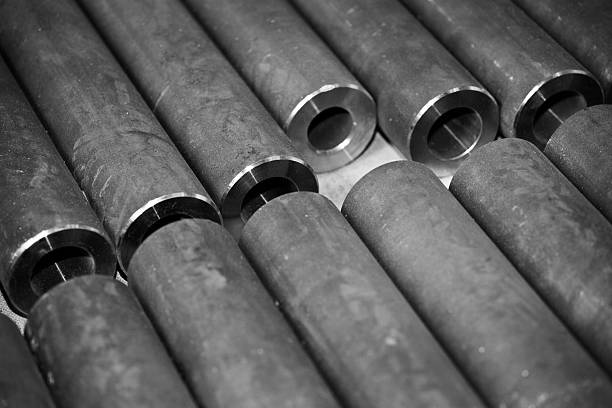
Some other orthopedic conditions like joint replacements could require implants made of metal which help to hold bone in place of human bone, but they come with a question What can I do to receive an MRI today? Before we discuss "Can I conduct an MRI with implanted metal?" let's talk about what MRI is. As the name suggests, the type of examination connected to "magnetic field". The goal of this type of examination is to trigger hydrogen protons inside the body by sending pulses through a variety different devices that cause them to vibrate. These resonance signals are later interpreted by computers to convert the signals into images. This seemingly straightforward work requires the use of a magnetic field. It must be strong. In the present, titanium alloy has become the most popular implant metal because of its low weight, high corrosion resistance, good biocompatibility with no magnetic characteristics, then are titanium alloy implants MRI safe? While titanium bar implants are generally safe, there are questions about their safety in MRI. For one, titanium is a nonmagnetic substance, which means it is able to be scanned without creating magnetic fields. One concern is the possibility of metal objects being removed in MRI procedures. This is because titanium isn't magnetic and can be scanned without causing magnetic fields. But, it's possible for other materials to be a danger to. This article focuses on the issues surrounding MRI safety, artifacts, CT protocol and reproducibility analysis. Read on to learn more about the titanium tool set safety and MRI scan security for titanium implants. Magnetic artifacts may create artificial signal variations and distorted object geometry in MRI images. They can also create large susceptibility gradients between an implant and surrounding tissues. These effects can even cause death-threatening injuries to medical devices which are ferromagnetic. The risk of heat is minimal in the position of a patient lying down, particularly if they are lying on their backs. While the artifacts in titanium MRI images are not significant in relation to their dimensions, the more likely they could affect clinical decision-making. For this reason, dedicated sequences of reduction of metal artifacts are being developed to reduce these impact. However, these techniques do not work as well on smaller artifactsthat aren't easily visible because of soft tissue. These findings were based on studies of regular tetrahedron-shaped square cubic and spherical titanium items. Materials were anisotropic and isotropic. The samples were placed on a nickel doped agarose gelphantom. Then, they were covered with the nickel nitrate hexahydrate. Three-Tela MR images of the samples were taken by using gradient echo sequences following they were placed on the phantom. The volume of the sample was calculated and subtracted from background to determine the quantity of artifacts present in the sample. The volume of artifacts grew in proportion to the normalized projection area of the sample. A CBCT and MRI protocol for titanium powder implants was created to assess the artifacts that are associated with these materials. The purpose of the study was to evaluate the volume of artifacts with respect to the implant's size and also to correlate these findings to exposure factors. Implants were embedded with ultrasound gel. A variety of settings were utilized to obtain MRI as well as CT images. The volume of artifacts was calculated by dividing it into a percentage of implant's volume.MR images of pilons containing SS screws showed streaks. These streaks penetrated the talar dome and were thought to be different from artifacts that exhibit susceptibility to titanium. These findings were further supported by the finding that the SS was inappropriate for MRI of the pilon. Furthermore the SS was of lower contrast to MRI for assessing the articular reduction of the pilon. Reproducibility analysis of MRI using titanium alloy is a crucial component in understanding the images. The volume contribution of titanium alloy mesh implants can vary significantly from one image. The volume content of titanium alloy mesh implants particularly can be erratic which makes a visual examination of attenuation maps crucial to identify signal voids. Utilizing a single slice titanium alloy scanner researchers examined a variety of titanium alloy MRI images to find out if the MRI image quality was degraded and if the method had any limitations. In one study, researchers employed titanium phantoms for three trepanation holes. Implants can be MRI-conditioned up to 3.0 Tesla and have been found to reduce the reproducibility of MR images. The susceptibility artifacts created by implants made of metal can exceed 5mm on an MRI T1. Because of the signal void and the lack of signal void, the authors decided to remove one repeat examination from both the group and individual analysis. The titanium bar that is employed in MRI tools, has minimal interference with the magnetic fields that are generated by MRI. There are disadvantages to consider prior to deciding whether or not you want to use these tools. MRI machines might not function correctly if they have titanium or other magnetic materials and patients with these metal implants should be evaluated carefully prior to having an MRI. Titanium alloys are less likely to create image distortion. In addition the alloys utilized in MRI are more durable than other materials. Implants for orthopedics, particularly those designed for the spine are made mostly of titanium or titanium. Titanium isn't affected by magnetic fields, and it does not move within the way of magnetic fields. Therefore, patients who have titanium implants are safe to undergo MRI. However, sometimes they may or may not interfere with the MRI image. Patients with spinal disorders or spinal internal fixation might benefit from this information.
0 Comments
Read More
Leave a Reply. |
 RSS Feed
